Copyright
©The Author(s) 2016.
World J Gastroenterol. Dec 14, 2016; 22(46): 10260-10266
Published online Dec 14, 2016. doi: 10.3748/wjg.v22.i46.10260
Published online Dec 14, 2016. doi: 10.3748/wjg.v22.i46.10260
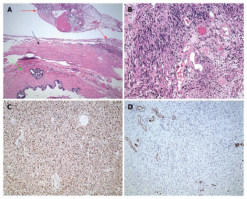
Figure 3 Microscopic examination and immunohistochemical staining.
A: Microscopically, the tumor (red arrow) with a capsule (black arrow) was adjacent to the cholecystic duct (green arrow) (HE, × 200); B: The tumor mainly consisted of spindle-shaped cells with both hypercellular and hypocellular areas (HE, × 200). Immunohistochemical investigation showed that the tumor was positive for protein S-100 (C) and negative for CD34 (D) (HE, × 100). HE: Hematoxylin and eosin.
- Citation: Xu SY, Sun K, Xie HY, Zhou L, Zheng SS, Wang WL. Schwannoma in the hepatoduodenal ligament: A case report and literature review. World J Gastroenterol 2016; 22(46): 10260-10266
- URL: https://www.wjgnet.com/1007-9327/full/v22/i46/10260.htm
- DOI: https://dx.doi.org/10.3748/wjg.v22.i46.10260


