Copyright
©The Author(s) 2016.
World J Gastroenterol. Dec 14, 2016; 22(46): 10198-10209
Published online Dec 14, 2016. doi: 10.3748/wjg.v22.i46.10198
Published online Dec 14, 2016. doi: 10.3748/wjg.v22.i46.10198
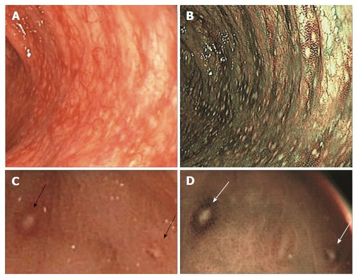
Figure 4 Endoscopic features of nodular lymphoid hyperplasia with red ring sign, due to hypervascularization at the base of the follicles, associated with granulocyte infiltrate.
A and B: A typical case of nodular lymphoid hyperplasia (NLH) with red ring sign (RRS) and diffuse macroscopic inflammation, in white light (WL) and narrow band imaging (NBI), respectively: RRS appears as a red (WL) or brown (NBI) outline surrounding NLH foci; C and D: A particular of two NLH foci with RRS, in WL and NBI, respectively (black and white arrows).
- Citation: Piscaglia AC, Laterza L, Cesario V, Gerardi V, Landi R, Lopetuso LR, Calò G, Fabbretti G, Brisigotti M, Stefanelli ML, Gasbarrini A. Nodular lymphoid hyperplasia: A marker of low-grade inflammation in irritable bowel syndrome? World J Gastroenterol 2016; 22(46): 10198-10209
- URL: https://www.wjgnet.com/1007-9327/full/v22/i46/10198.htm
- DOI: https://dx.doi.org/10.3748/wjg.v22.i46.10198


