Copyright
©The Author(s) 2016.
World J Gastroenterol. Dec 14, 2016; 22(46): 10198-10209
Published online Dec 14, 2016. doi: 10.3748/wjg.v22.i46.10198
Published online Dec 14, 2016. doi: 10.3748/wjg.v22.i46.10198
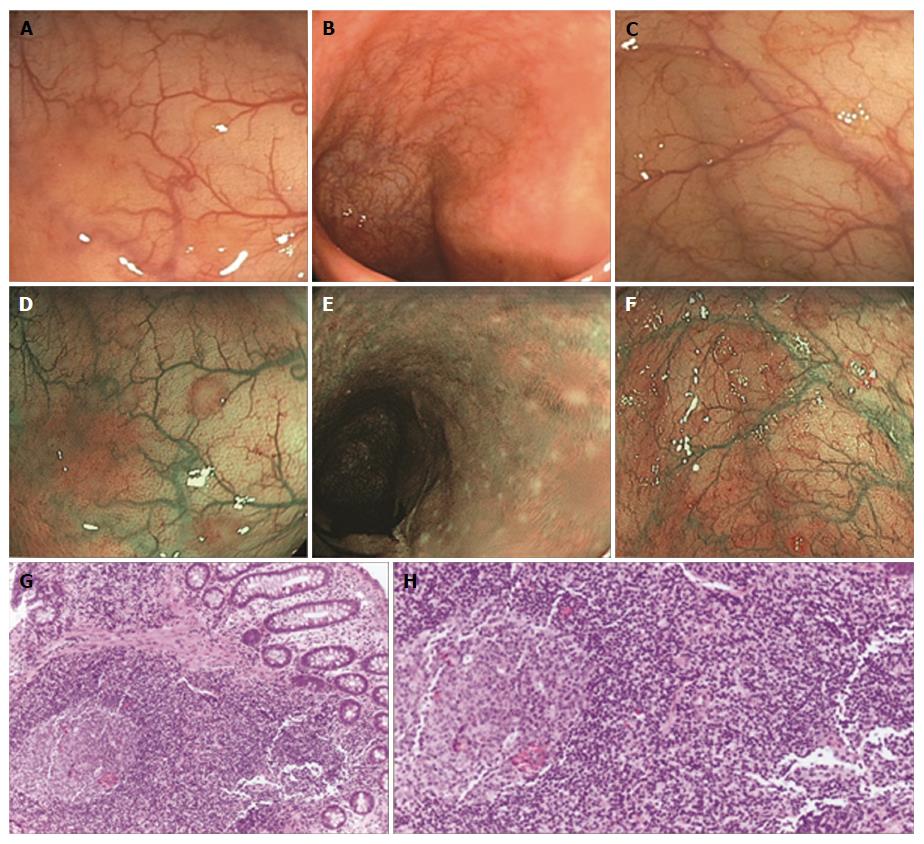
Figure 2 Endoscopic and histological features of nodular lymphoid hyperplasia.
A-F: Three typical cases of nodular lymphoid hyperplasia (NLH), as observed at white light (WL) standard endoscopy (A-C) and narrow band imaging (NBI) endoscopy (D-F), respectively. NLH appear as slightly raised whitish areas, usually < 5 mm in diameter, closely spaced, difficult to recognize at WL, and easier to observe by NBI; G and H: Show the histological appearance of NLH (hematoxylin-eosin staining), as clusters of ≤ 10 lymphoid nodules, composed of hyperplastic benign lymphoid tissue.
- Citation: Piscaglia AC, Laterza L, Cesario V, Gerardi V, Landi R, Lopetuso LR, Calò G, Fabbretti G, Brisigotti M, Stefanelli ML, Gasbarrini A. Nodular lymphoid hyperplasia: A marker of low-grade inflammation in irritable bowel syndrome? World J Gastroenterol 2016; 22(46): 10198-10209
- URL: https://www.wjgnet.com/1007-9327/full/v22/i46/10198.htm
- DOI: https://dx.doi.org/10.3748/wjg.v22.i46.10198


