Copyright
©The Author(s) 2016.
World J Gastroenterol. Dec 7, 2016; 22(45): 9880-9897
Published online Dec 7, 2016. doi: 10.3748/wjg.v22.i45.9880
Published online Dec 7, 2016. doi: 10.3748/wjg.v22.i45.9880
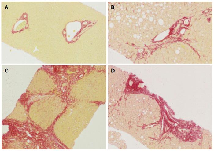
Figure 1 Histology of normal liver, fibrosis and cirrhosis.
A: Representative histological images (using Sirius red staining), normal liver; B: Mild to moderate fibrosis with portal tract expansion (METAVIR F = 2, Ishak stage 3); C: Moderate “bridging” fibrosis (METAVIR F = 3, Ishak stage 4); D: Cirrhosis (METAVIR F = 4, Ishak 5 or 6).
- Citation: Karanjia RN, Crossey MME, Cox IJ, Fye HKS, Njie R, Goldin RD, Taylor-Robinson SD. Hepatic steatosis and fibrosis: Non-invasive assessment. World J Gastroenterol 2016; 22(45): 9880-9897
- URL: https://www.wjgnet.com/1007-9327/full/v22/i45/9880.htm
- DOI: https://dx.doi.org/10.3748/wjg.v22.i45.9880


