Copyright
©The Author(s) 2016.
World J Gastroenterol. Nov 28, 2016; 22(44): 9822-9828
Published online Nov 28, 2016. doi: 10.3748/wjg.v22.i44.9822
Published online Nov 28, 2016. doi: 10.3748/wjg.v22.i44.9822
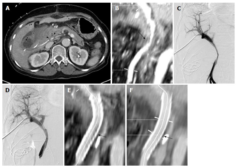
Figure 3 A 68-year-old woman (patient No.
8) presenting with main portal vein stenosis after pylorus-preserving pancreatoduodenectomy for duodenal gastrointestinal stromal tumor. She presented with anorexia and increased JP drain output during the follow-up period. A: Axial CT image 13 d after surgery showing stenosis at junction between the PV and SMV (arrowheads); B: Curved planar reformatted (CPR) image from abdominal CT 13 d after surgery showing stenosis at junction between the PV and SMV (arrow); C: Percutaneous transhepatic portogram showing severe stenosis (> 50%) at the PV-SMV junction. The pressure gradient was not measured because of definite stagnation of the contrast medium. The contrast medium injected at the distal SMV is clearly stagnant; D: Portogram showing metallic stent in the main PV and SMV and the elimination of stenosis. After the procedure, the patient’s symptoms and signs improved; E: CPR image from abdominal CT 2 d after stenting showing patent stent with small in-stent low-density area (arrow); F: The extent of small in-stent low-density area decreased on CPR image from abdominal CT scan 555 d after stenting (arrow). CT: Computed tomography; PV: Portal vein; SMV: Superior mesenteric vein.
- Citation: Jeon UB, Kim CW, Kim TU, Choo KS, Jang JY, Nam KJ, Chu CW, Ryu JH. Therapeutic efficacy and stent patency of transhepatic portal vein stenting after surgery. World J Gastroenterol 2016; 22(44): 9822-9828
- URL: https://www.wjgnet.com/1007-9327/full/v22/i44/9822.htm
- DOI: https://dx.doi.org/10.3748/wjg.v22.i44.9822


