Copyright
©The Author(s) 2016.
World J Gastroenterol. Nov 28, 2016; 22(44): 9765-9774
Published online Nov 28, 2016. doi: 10.3748/wjg.v22.i44.9765
Published online Nov 28, 2016. doi: 10.3748/wjg.v22.i44.9765
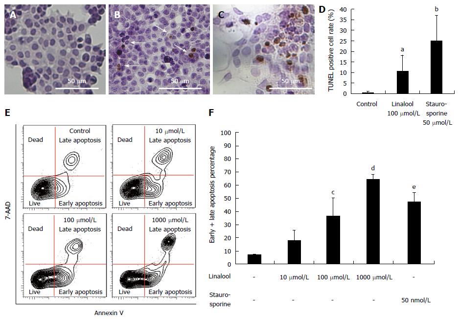
Figure 2 Anticancer mechanism of linalool.
A: TUNEL staining of HCT 116 cells in the control group; B: Staining of HCT 116 cells treated with 100 μmol/L linalool; C: Staining of HCT 116 cells treated with 50 nmol/L staurosporine. The corresponding percentage of TUNEL-positive cells (white arrow) was 0.7% ± 0.7% (A), 10.5% ± 7.7% (B), and 25.1% ± 11.2% (C); D: The percentage of apoptotic cells in each group (mean ± SD, n = 10). aP < 0.05 vs control group; bP < 0.05 vs 100 μmol/L linalool group; E: Flow cytometry using Annexin V for the detection of live cells, cells showing early and late apoptosis, and cell death; F: Percentages of early and late apoptosis fraction. cP < 0.05 vs control group; dP < 0.01 vs control group; eP < 0.05 vs control group; one-way ANOVA followed by Dunnett’s test, n = 3.
- Citation: Iwasaki K, Zheng YW, Murata S, Ito H, Nakayama K, Kurokawa T, Sano N, Nowatari T, Villareal MO, Nagano YN, Isoda H, Matsui H, Ohkohchi N. Anticancer effect of linalool via cancer-specific hydroxyl radical generation in human colon cancer. World J Gastroenterol 2016; 22(44): 9765-9774
- URL: https://www.wjgnet.com/1007-9327/full/v22/i44/9765.htm
- DOI: https://dx.doi.org/10.3748/wjg.v22.i44.9765


