Copyright
©The Author(s) 2016.
World J Gastroenterol. Nov 21, 2016; 22(43): 9654-9660
Published online Nov 21, 2016. doi: 10.3748/wjg.v22.i43.9654
Published online Nov 21, 2016. doi: 10.3748/wjg.v22.i43.9654
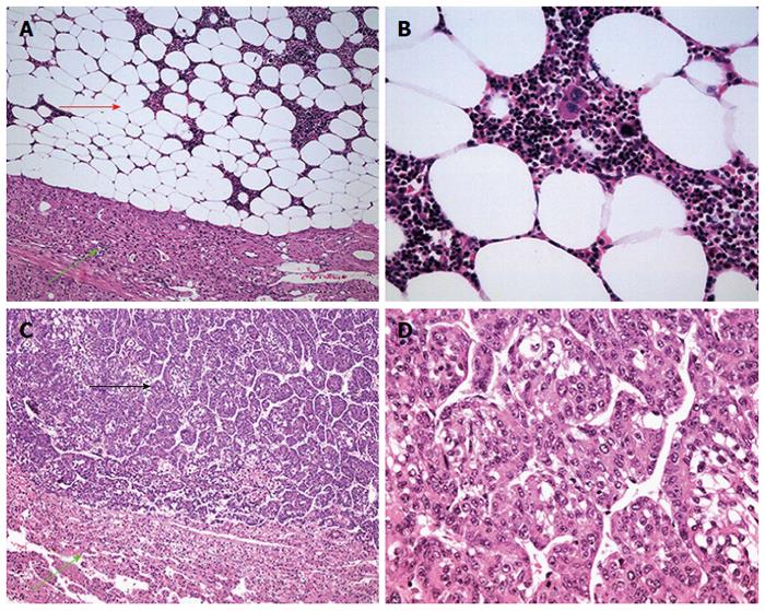
Figure 2 Microscopic examination.
A and B: The tumor (red arrow) in the right liver lobe consists of adipose tissue and hematopoietic elements containing erythroid tissue, megakaryocytes and myeloid colonies, consistent with a myelolipoma (A: HE, 100 ×; B: HE, 400 ×); C and D: The tumor cells (black arrow) in the left liver lobe are in a funicular arrangement and are growing invasively with significant atypia, compatible with a malignant hepatic cancer (C: HE, 100 ×; D: HE, 400 ×). The areas marked by green arrows are normal liver tissue. HE: Hematoxylin and eosin.
- Citation: Xu SY, Xie HY, Zhou L, Zheng SS, Wang WL. Synchronous occurrence of a hepatic myelolipoma and two hepatocellular carcinomas. World J Gastroenterol 2016; 22(43): 9654-9660
- URL: https://www.wjgnet.com/1007-9327/full/v22/i43/9654.htm
- DOI: https://dx.doi.org/10.3748/wjg.v22.i43.9654


