Copyright
©The Author(s) 2016.
World J Gastroenterol. Nov 21, 2016; 22(43): 9525-9533
Published online Nov 21, 2016. doi: 10.3748/wjg.v22.i43.9525
Published online Nov 21, 2016. doi: 10.3748/wjg.v22.i43.9525
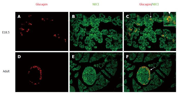
Figure 4 Localization of the NBC1 and glucagon determined by immunofluorescence detection in pancreas of embryonic day 18.
5 and adult rat. Labeling by the NBC1 antibodies was detected with an FITC (green)-labeled secondary antibody. Labeling of glucagon was detected with a CY3 (red)-labeled secondary antibody on the same section. The overlap of NBC1 (green) and glucagon (red) labeling creates orange color. NBC1 positive cells and glucagon positive cells can be detected in two pancreas sections tested. Obviously, the two proteins are predominantly colocalized in embryonic day (E)18.5 and adult rat (arrows). All of the primary magnifications are × 400. Insets, higher magnifications of the areas indicated by arrow in E18.5.
- Citation: Cao LH, Xia CC, Shi ZC, Wang N, Gu ZH, Yu LZ, Wan Q, De W. Na+/HCO3- cotransporter is expressed on β and α cells during rat pancreatic development. World J Gastroenterol 2016; 22(43): 9525-9533
- URL: https://www.wjgnet.com/1007-9327/full/v22/i43/9525.htm
- DOI: https://dx.doi.org/10.3748/wjg.v22.i43.9525


