Copyright
©The Author(s) 2016.
World J Gastroenterol. Nov 14, 2016; 22(42): 9445-9450
Published online Nov 14, 2016. doi: 10.3748/wjg.v22.i42.9445
Published online Nov 14, 2016. doi: 10.3748/wjg.v22.i42.9445
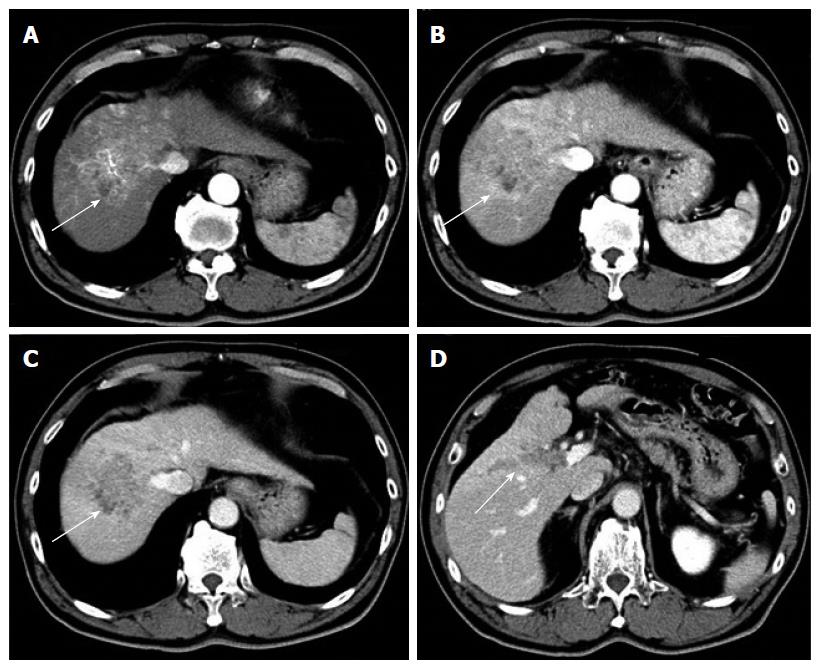
Figure 1 Contrast-enhanced computed tomography before sorafenib introduction.
Heterogeneous hypervascularized tumor (A-C, arrow) in the right paramedian sector, showing early enhancement in the arterial phase and wash-out in the late phase together with portal vein tumor thrombosis (D, arrow) limited to the first-order branch and invading the right portal vein.
- Citation: Takano M, Kokudo T, Miyazaki Y, Kageyama Y, Takahashi A, Amikura K, Sakamoto H. Complete response with sorafenib and transcatheter arterial chemoembolization in unresectable hepatocellular carcinoma. World J Gastroenterol 2016; 22(42): 9445-9450
- URL: https://www.wjgnet.com/1007-9327/full/v22/i42/9445.htm
- DOI: https://dx.doi.org/10.3748/wjg.v22.i42.9445


