Copyright
©The Author(s) 2016.
World J Gastroenterol. Nov 14, 2016; 22(42): 9419-9426
Published online Nov 14, 2016. doi: 10.3748/wjg.v22.i42.9419
Published online Nov 14, 2016. doi: 10.3748/wjg.v22.i42.9419
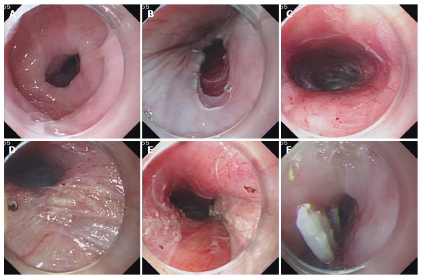
Figure 2 Case illustration of peroral endoscopic full-thickness myotomy.
A: Endoscopy showing dilated esophagus; B: Longitudinal mucosal incision was made to create a tunnel entry; C: Submucosal tunnel; D: Circular myotomy was initially performed; E: Full-thickness myotomy was performed; F: Tunnel entry was closed with several clips.
- Citation: Wang XH, Tan YY, Zhu HY, Li CJ, Liu DL. Full-thickness myotomy is associated with higher rate of postoperative gastroesophageal reflux disease. World J Gastroenterol 2016; 22(42): 9419-9426
- URL: https://www.wjgnet.com/1007-9327/full/v22/i42/9419.htm
- DOI: https://dx.doi.org/10.3748/wjg.v22.i42.9419


