Copyright
©The Author(s) 2016.
World J Gastroenterol. Nov 14, 2016; 22(42): 9324-9332
Published online Nov 14, 2016. doi: 10.3748/wjg.v22.i42.9324
Published online Nov 14, 2016. doi: 10.3748/wjg.v22.i42.9324
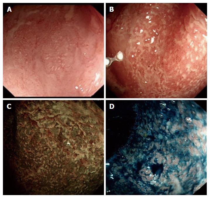
Figure 1 Assessment of inflamed colon with white light endoscopy, narrow band imaging and chromoendoscopy.
A: White light assessment with standard definition endosocpe reveals areas with superficial ulceration interspersed with areas of patchy obliteration of mucosal vascular pattern; B: High resolution endoscope allows more detailed assessment including crypt openings and disrupted vascular architecture; C: NBI assessment of moderately active UC shows obscured vascular pattern; white mucosal spots which represent mucous exudates giving the characteristic appearance of “Coral reaf” like mucosa; D: Chromoendoscopy shows the mucosal damage with disruption of pit pattern and complete destruction of vascular pattern. Ulcer margins are seen more prominent with contrast enhancement.
- Citation: Mohammed N, Subramanian V. Clinical relevance of endoscopic assessment of inflammation in ulcerative colitis: Can endoscopic evaluation predict outcomes? World J Gastroenterol 2016; 22(42): 9324-9332
- URL: https://www.wjgnet.com/1007-9327/full/v22/i42/9324.htm
- DOI: https://dx.doi.org/10.3748/wjg.v22.i42.9324


