Copyright
©The Author(s) 2016.
World J Gastroenterol. Nov 7, 2016; 22(41): 9242-9246
Published online Nov 7, 2016. doi: 10.3748/wjg.v22.i41.9242
Published online Nov 7, 2016. doi: 10.3748/wjg.v22.i41.9242
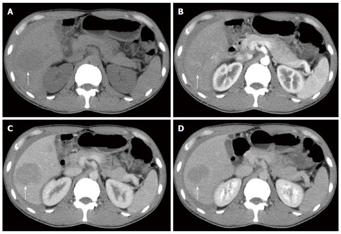
Figure 1 Contrast-enhanced computed tomography performed two years ago demonstrated multiple nodules at the right lobe of the liver.
A: Non-contrast-enhanced computed tomography scan; B: Arterial phase; C: Portal phase; and D: Venous phase.
- Citation: Hao B, Guo W, Luo NN, Fu H, Chen HJ, Zhao L, Wu H, Sun L. Metabolic imaging for guidance of curative treatment of isolated pelvic implantation metastasis after resection of spontaneously ruptured hepatocellular carcinoma: A case report. World J Gastroenterol 2016; 22(41): 9242-9246
- URL: https://www.wjgnet.com/1007-9327/full/v22/i41/9242.htm
- DOI: https://dx.doi.org/10.3748/wjg.v22.i41.9242


