Copyright
©The Author(s) 2016.
World J Gastroenterol. Nov 7, 2016; 22(41): 9235-9241
Published online Nov 7, 2016. doi: 10.3748/wjg.v22.i41.9235
Published online Nov 7, 2016. doi: 10.3748/wjg.v22.i41.9235
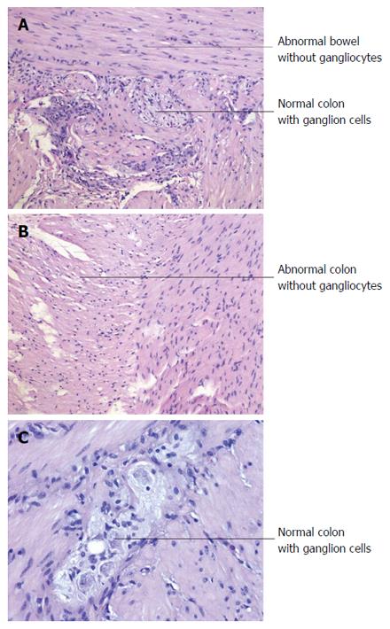
Figure 3 Postsurgical pathological examination.
A: Shows the junction of the normal and the abnormal bowel contained in the resected specimen (magnification × 100); B: Indicates the resected abnormal bowel segment without ganglion cells (magnification × 100). The cutting edge contained abundant normal ganglion cells (C, magnification × 200), ensuring the total and complete removal of the abnormal and aganglioniccolon segment.
- Citation: Wei ZJ, Huang L, Xu AM. Reoperation in an adult female with "right-sided" Hirschsprung's disease complicated by refractory hypertension and cough. World J Gastroenterol 2016; 22(41): 9235-9241
- URL: https://www.wjgnet.com/1007-9327/full/v22/i41/9235.htm
- DOI: https://dx.doi.org/10.3748/wjg.v22.i41.9235


