Copyright
©The Author(s) 2016.
World J Gastroenterol. Nov 7, 2016; 22(41): 9229-9234
Published online Nov 7, 2016. doi: 10.3748/wjg.v22.i41.9229
Published online Nov 7, 2016. doi: 10.3748/wjg.v22.i41.9229
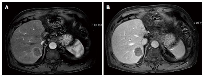
Figure 3 Magnetic resonance imaging of the liver after 6 mo.
Five nodules were detected in the right lobe. The biggest was 3.3 cm. The nodules showed rim enhancement on the arterial phase (A) and low density on the portal phase (B).
- Citation: Choi GH, Ann SY, Lee SI, Kim SB, Song IH. Collision tumor of hepatocellular carcinoma and neuroendocrine carcinoma involving the liver: Case report and review of the literature. World J Gastroenterol 2016; 22(41): 9229-9234
- URL: https://www.wjgnet.com/1007-9327/full/v22/i41/9229.htm
- DOI: https://dx.doi.org/10.3748/wjg.v22.i41.9229


