Copyright
©The Author(s) 2016.
World J Gastroenterol. Nov 7, 2016; 22(41): 9229-9234
Published online Nov 7, 2016. doi: 10.3748/wjg.v22.i41.9229
Published online Nov 7, 2016. doi: 10.3748/wjg.v22.i41.9229
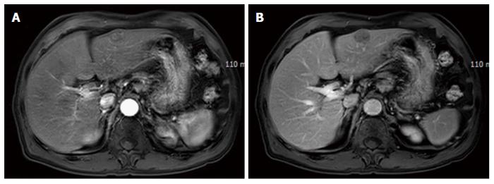
Figure 1 Magnetic resonance imaging of the liver.
A 2.2 cm × 2.2 cm sized lobular contoured mass was found on segment 3. It showed mild enhancement in the arterial phase (A) and a washed-out pattern in the portal phase (B).
- Citation: Choi GH, Ann SY, Lee SI, Kim SB, Song IH. Collision tumor of hepatocellular carcinoma and neuroendocrine carcinoma involving the liver: Case report and review of the literature. World J Gastroenterol 2016; 22(41): 9229-9234
- URL: https://www.wjgnet.com/1007-9327/full/v22/i41/9229.htm
- DOI: https://dx.doi.org/10.3748/wjg.v22.i41.9229


