Copyright
©The Author(s) 2016.
World J Gastroenterol. Nov 7, 2016; 22(41): 9196-9204
Published online Nov 7, 2016. doi: 10.3748/wjg.v22.i41.9196
Published online Nov 7, 2016. doi: 10.3748/wjg.v22.i41.9196
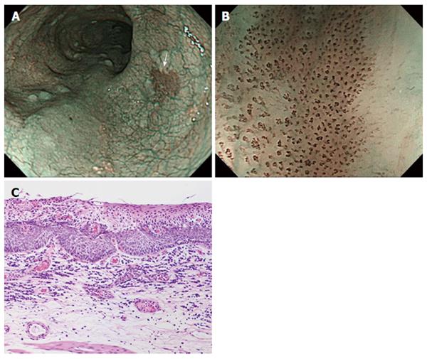
Figure 3 Representative case of superficial squamous cell carcinoma.
A: On non-magnifying NBI endoscopy, the lesion demonstrated a well-demarcated brownish area; B: The lesion has all six of the diagnostic findings obtained by using NBI-ME; C: Histology from endoscopic submucosal dissection showing squamous cell carcinoma invading up to the lamina propria mucosae. NBI: Narrow Band Imaging; NBI-ME: Narrow Band Imaging combined with magnifying endoscopy.
- Citation: Dobashi A, Goda K, Yoshimura N, Ohya TR, Kato M, Sumiyama K, Matsushima M, Hirooka S, Ikegami M, Tajiri H. Simplified criteria for diagnosing superficial esophageal squamous neoplasms using Narrow Band Imaging magnifying endoscopy. World J Gastroenterol 2016; 22(41): 9196-9204
- URL: https://www.wjgnet.com/1007-9327/full/v22/i41/9196.htm
- DOI: https://dx.doi.org/10.3748/wjg.v22.i41.9196


