Copyright
©The Author(s) 2016.
World J Gastroenterol. Oct 28, 2016; 22(40): 9028-9034
Published online Oct 28, 2016. doi: 10.3748/wjg.v22.i40.9028
Published online Oct 28, 2016. doi: 10.3748/wjg.v22.i40.9028
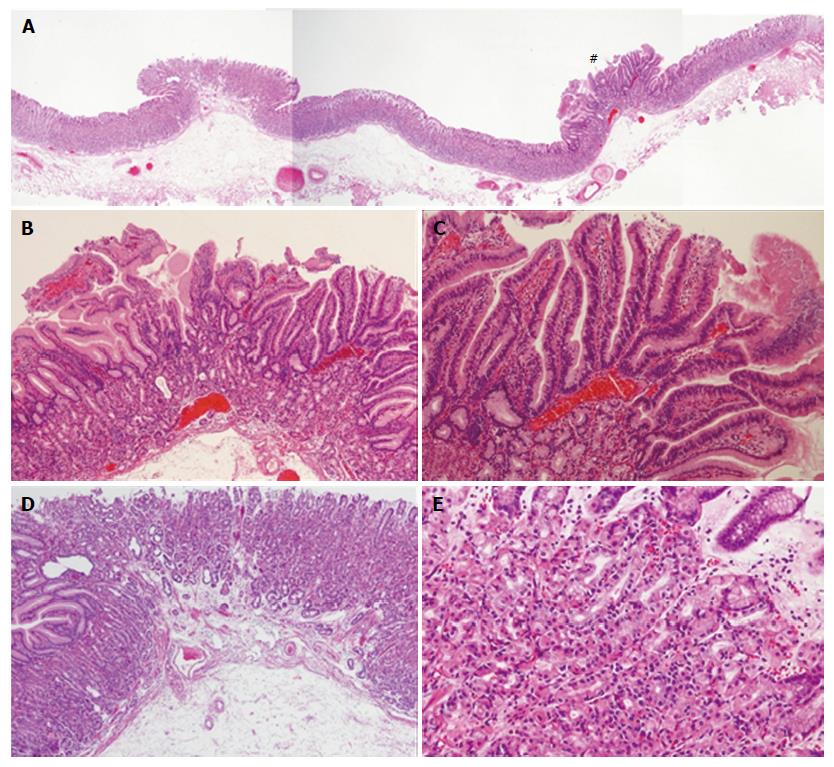
Figure 4 Histological examination of the endoscopic submucosal dissection specimen from Case 2.
Shows that the right side lesion (#) is consistent with reddish polypoid and the left side lesion is consistent with isochromatic small polyp (A). High magnification of the right side lesion (#) shows irregularly branching tumor glands with atypical nuclei at the surface part of the lesion, and proliferation of fundic glands with some cystic dilatation at the basal part of the lesion, which was diagnosed as very well-differentiated adenocarcinoma occurring in fundic gland polyps (FGPs) (B and C). High magnification of the left side lesion shows proliferation of fundic glands with some cystic dilatation, which was diagnosed as FGP without dysplasia (D and E). FGP: Gastric fundic gland polyp.
- Citation: Togo K, Ueo T, Yonemasu H, Honda H, Ishida T, Tanabe H, Yao K, Iwashita A, Murakami K. Two cases of adenocarcinoma occurring in sporadic fundic gland polyps observed by magnifying endoscopy with narrow band imaging. World J Gastroenterol 2016; 22(40): 9028-9034
- URL: https://www.wjgnet.com/1007-9327/full/v22/i40/9028.htm
- DOI: https://dx.doi.org/10.3748/wjg.v22.i40.9028


