Copyright
©The Author(s) 2016.
World J Gastroenterol. Jan 28, 2016; 22(4): 1607-1616
Published online Jan 28, 2016. doi: 10.3748/wjg.v22.i4.1607
Published online Jan 28, 2016. doi: 10.3748/wjg.v22.i4.1607
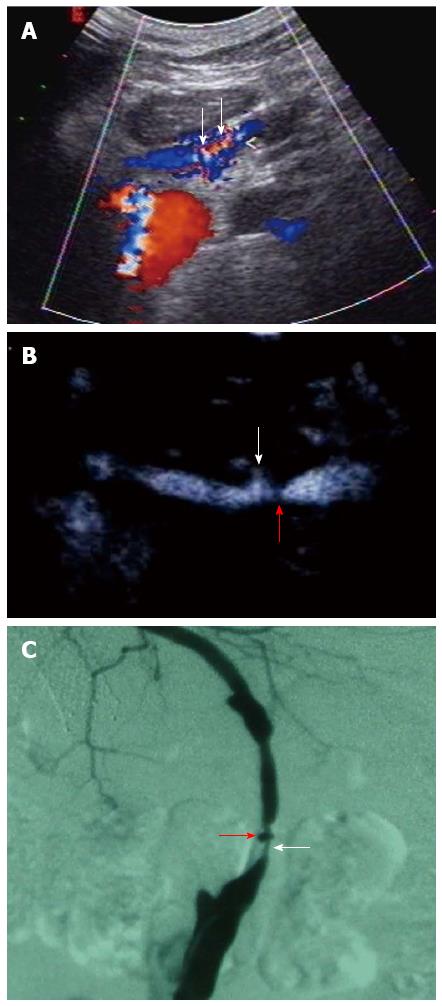
Figure 3 Hepatic artery stenosis and pseudo-aneurysm.
A 43-year-old recipient with arterial bypass arising from the infrarenal aorta through the transverse mesocolon with an iliac artery graft tunneled. A: Color Doppler ultrasound shows turbulent flow of the graft artery (arrows); B: Contrast-enhanced ultrasound shows stenosis at the graft artery (arrows) and continuing patency of the pseudo-aneurysm with adjacent site of stenosis (red arrow); C: Conventional angiography confirms hepatic artery stenosis (arrow) and pseudo-aneurysm (red arrow).
- Citation: Ren J, Wu T, Zheng BW, Tan YY, Zheng RQ, Chen GH. Application of contrast-enhanced ultrasound after liver transplantation: Current status and perspectives. World J Gastroenterol 2016; 22(4): 1607-1616
- URL: https://www.wjgnet.com/1007-9327/full/v22/i4/1607.htm
- DOI: https://dx.doi.org/10.3748/wjg.v22.i4.1607


