Copyright
©The Author(s) 2016.
World J Gastroenterol. Jan 28, 2016; 22(4): 1607-1616
Published online Jan 28, 2016. doi: 10.3748/wjg.v22.i4.1607
Published online Jan 28, 2016. doi: 10.3748/wjg.v22.i4.1607
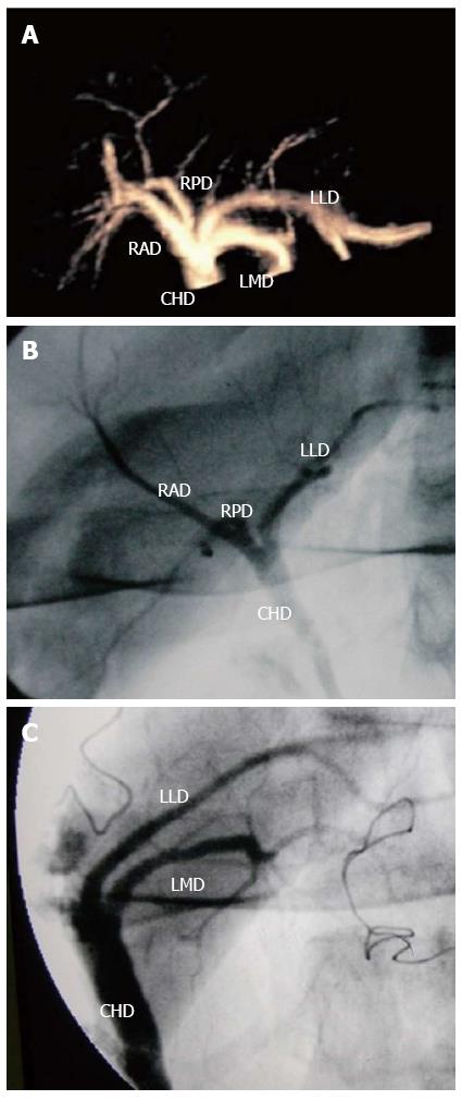
Figure 1 Three-dimensional contrast-enhanced ultrasonic cholangiography for displaying the biliary tree.
A 21-year-old living donor liver with normal biliary anatomy. A: On the anterior-posterior 3D contrast-enhanced ultrasonic cholangiography image delineating the biliary system, the common hepatic duct (CHD), right posterior duct (RPD), right anterior duct (RAD), left lateral duct (LLD) and left median duct (LMD) are well seen; B: IOC image also shows CHD, RPD, RAD and LLD before hepatectomy; C: IOC image shows CHD, LLD and LMD after hepatectomy. IOC: intraoperative cholangiography.
- Citation: Ren J, Wu T, Zheng BW, Tan YY, Zheng RQ, Chen GH. Application of contrast-enhanced ultrasound after liver transplantation: Current status and perspectives. World J Gastroenterol 2016; 22(4): 1607-1616
- URL: https://www.wjgnet.com/1007-9327/full/v22/i4/1607.htm
- DOI: https://dx.doi.org/10.3748/wjg.v22.i4.1607


