Copyright
©The Author(s) 2016.
World J Gastroenterol. Oct 21, 2016; 22(39): 8844-8848
Published online Oct 21, 2016. doi: 10.3748/wjg.v22.i39.8844
Published online Oct 21, 2016. doi: 10.3748/wjg.v22.i39.8844
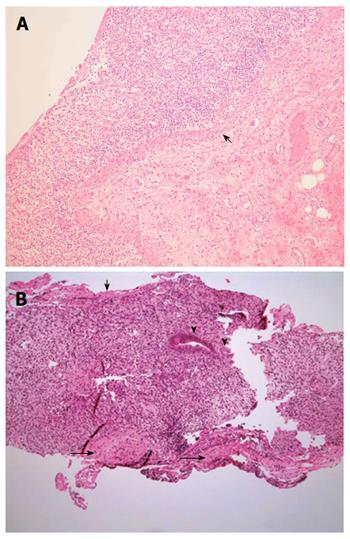
Figure 3 Post-mortem histopathology.
A: Small bowel. The high power view of small bowel shows focal full thickness mucosal ulceration with inflamed granulation tissue and preservation of the muscularis mucosae (arrow); B: Colon. Endoscopic biopsy of the left colon approximately one month after commencing cyclophosphamide showed surface fibrin exudate (short arrow), severe mucosal ulceration and granulation tissue with a few residual glands (arrowheads). There were no viral inclusions or granulomas. Preserved muscularis mucosae is seen (long arrows). Hematoxylin-eosin staining, magnification × 100.
- Citation: Yang LS, Cameron K, Papaluca T, Basnayake C, Jackett L, McKelvie P, Goodman D, Demediuk B, Bell SJ, Thompson AJ. Cyclophosphamide-associated enteritis: A rare association with severe enteritis. World J Gastroenterol 2016; 22(39): 8844-8848
- URL: https://www.wjgnet.com/1007-9327/full/v22/i39/8844.htm
- DOI: https://dx.doi.org/10.3748/wjg.v22.i39.8844


