Copyright
©The Author(s) 2016.
World J Gastroenterol. Oct 21, 2016; 22(39): 8820-8830
Published online Oct 21, 2016. doi: 10.3748/wjg.v22.i39.8820
Published online Oct 21, 2016. doi: 10.3748/wjg.v22.i39.8820
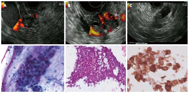
Figure 2 Endoscopic ultrasound and cytology images from various lesions.
A: Adenocarcinoma of the pancreatic head. Cytology shows a cluster of carcinoma cells with crowding and nuclear pleomorphism with anisokaryosis and prominent nucleoli (Pap test); B: Neuroendocrine tumors (insulinoma) of the pancreas Cytology shows a group of small monomorphic cells with uniform round nuclei and fine chromatin; C: Pancreatic metastasis of malignant melanoma: Cytology shows pleomorphic tumor cells with anisokaryosis and a high nuclear-to-cytoplasmic ratio. Positive staining for melanoma antigen recognized by T-cells-1.
- Citation: Sterlacci W, Sioulas AD, Veits L, Gönüllü P, Schachschal G, Groth S, Anders M, Kontos CK, Topalidis T, Hinsch A, Vieth M, Rösch T, Denzer UW. 22-gauge core vs 22-gauge aspiration needle for endoscopic ultrasound-guided sampling of abdominal masses. World J Gastroenterol 2016; 22(39): 8820-8830
- URL: https://www.wjgnet.com/1007-9327/full/v22/i39/8820.htm
- DOI: https://dx.doi.org/10.3748/wjg.v22.i39.8820


