Copyright
©The Author(s) 2016.
World J Gastroenterol. Oct 21, 2016; 22(39): 8779-8789
Published online Oct 21, 2016. doi: 10.3748/wjg.v22.i39.8779
Published online Oct 21, 2016. doi: 10.3748/wjg.v22.i39.8779
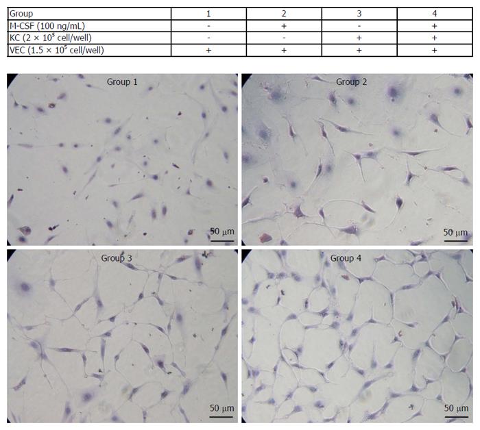
Figure 6 Proliferation of isolated vascular endothelial cells.
Isolated vascular endothelial cells (VECs) were co-cultured with or without the isolated Kupffer cells in media containing of macrophage colony-stimulating factor (M-CSF). Cell proliferation of VECs was determined as described in the Patients and Methods (n = 5). Treatments of each group are shown in the table. Representative photomicrographs are shown. Original magnification, × 400.
- Citation: Kono H, Fujii H, Furuya S, Hara M, Hirayama K, Akazawa Y, Nakata Y, Tsuchiya M, Hosomura N, Sun C. Macrophage colony-stimulating factor expressed in non-cancer tissues provides predictive powers for recurrence in hepatocellular carcinoma. World J Gastroenterol 2016; 22(39): 8779-8789
- URL: https://www.wjgnet.com/1007-9327/full/v22/i39/8779.htm
- DOI: https://dx.doi.org/10.3748/wjg.v22.i39.8779


