Copyright
©The Author(s) 2016.
World J Gastroenterol. Oct 14, 2016; 22(38): 8631-8637
Published online Oct 14, 2016. doi: 10.3748/wjg.v22.i38.8631
Published online Oct 14, 2016. doi: 10.3748/wjg.v22.i38.8631
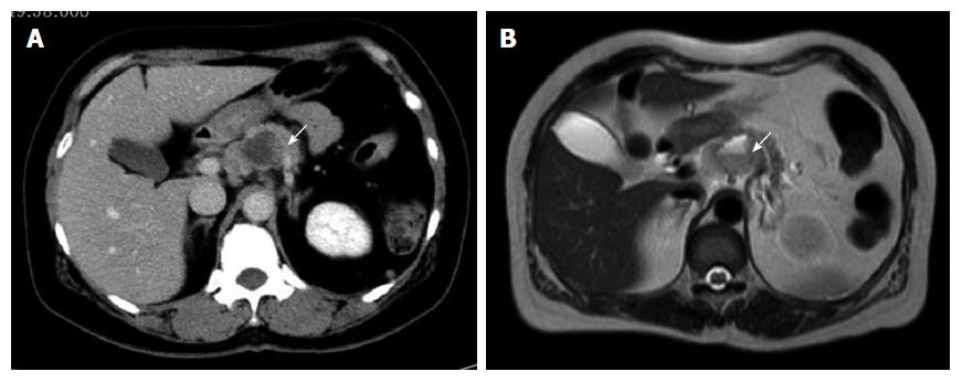
Figure 1 Imaging studies.
A: Abdominal computed tomography revealing a well-demarcated cystic tumor with slight enhancement of the peripheral portion of the lesion in the pancreatic body (arrow); B: Magnetic resonance imaging revealing mixed signal intensity of the tumor and a fluid-fluid level on T2-weighted imaging (arrow).
- Citation: Hoshimoto S, Matsui J, Miyata R, Takigawa Y, Miyauchi J. Anaplastic carcinoma of the pancreas: Case report and literature review of reported cases in Japan. World J Gastroenterol 2016; 22(38): 8631-8637
- URL: https://www.wjgnet.com/1007-9327/full/v22/i38/8631.htm
- DOI: https://dx.doi.org/10.3748/wjg.v22.i38.8631


