Copyright
©The Author(s) 2016.
World J Gastroenterol. Oct 14, 2016; 22(38): 8624-8630
Published online Oct 14, 2016. doi: 10.3748/wjg.v22.i38.8624
Published online Oct 14, 2016. doi: 10.3748/wjg.v22.i38.8624
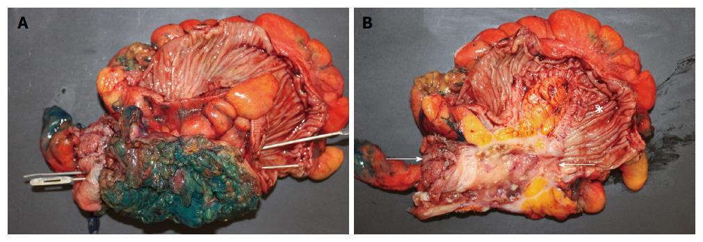
Figure 2 Open laparotomy revealed an extensive mass involving appendix, cecum, sigmoid colon, anterior abdominal wall, and urinary bladder.
A: Gross pathologic photograph shows entire surgically resected specimen (including appendix, cecum, sigmoid colon, part of bladder, and anterior abdominal wall) after bisecting the sigmoid colon that exposes the haustral folds within sigmoid, but before bisecting the centro-inferior tumor (note green ink identifying surgical margins of tumor). Insertion of two parallel probes (one inserted from left to right and the other inserted from right to left) demonstrate a fistula traveling from the cecum, on the bottom left, to the sigmoid, on the bottom right. Bisection of the centro-inferior tumor (B) reveals that the appendix forms the malignant fistula; B: Gross pathologic specimen after bisection of centro-inferior tumor (seen before bisection in A) exposes an irregular, cystically dilated, malignant appendix that forms the malignant fistula. It is lined by a variably thick wall composed of the mucinous adenocarcinoma and filled with viscous, mucoid, content. Sigmoid (star). Cecum is at left bottom of photograph immediately adjacent to arrow.
- Citation: Hakim S, Amin M, Cappell MS. Limited, local, extracolonic spread of mucinous appendiceal adenocarcinoma after perforation with formation of a malignant appendix-to-sigmoid fistula: Case report and literature review. World J Gastroenterol 2016; 22(38): 8624-8630
- URL: https://www.wjgnet.com/1007-9327/full/v22/i38/8624.htm
- DOI: https://dx.doi.org/10.3748/wjg.v22.i38.8624


