Copyright
©The Author(s) 2016.
World J Gastroenterol. Oct 14, 2016; 22(38): 8624-8630
Published online Oct 14, 2016. doi: 10.3748/wjg.v22.i38.8624
Published online Oct 14, 2016. doi: 10.3748/wjg.v22.i38.8624
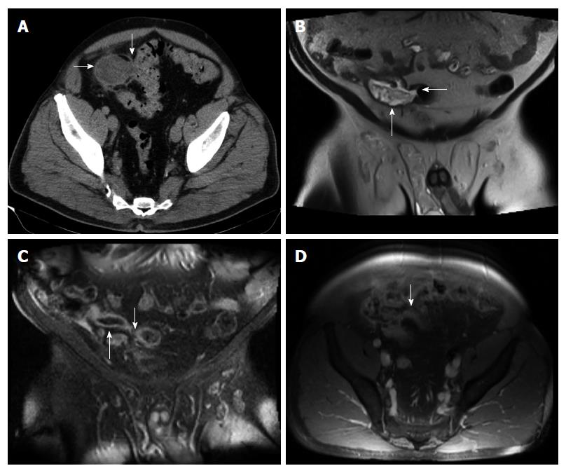
Figure 1 Abdomino-pelvic computed tomography.
A: Axial computed tomography (CT) image of the pelvis, enhanced with iodinated contrast, demonstrates a dilated, heterogeneous appendix (horizontal arrow) forming a fistula with sigmoid colon (fistulous connection at vertical arrow); B: Coronal T2-weighted fluid-sensitive MRI sequence depicts dilated, fluid-filled, appendix (vertical arrow), extending and forming a fistula with the sigmoid colon (horizontal arrow); C: Coronal post-gadolinium contrast enhanced T1-weighted MRI image demonstrates dilated appendix with non-enhanced, central fluid and enhanced wall (upward arrow) that extends towards sigmoid colon (downward arrow); D: Axial post-gadolinium contrast enhanced T1-weighted MRI image demonstrates abnormally thick and enhanced appendiceal wall (arrow) without significant surrounding inflammatory changes.
- Citation: Hakim S, Amin M, Cappell MS. Limited, local, extracolonic spread of mucinous appendiceal adenocarcinoma after perforation with formation of a malignant appendix-to-sigmoid fistula: Case report and literature review. World J Gastroenterol 2016; 22(38): 8624-8630
- URL: https://www.wjgnet.com/1007-9327/full/v22/i38/8624.htm
- DOI: https://dx.doi.org/10.3748/wjg.v22.i38.8624


