Copyright
©The Author(s) 2016.
World J Gastroenterol. Oct 14, 2016; 22(38): 8605-8614
Published online Oct 14, 2016. doi: 10.3748/wjg.v22.i38.8605
Published online Oct 14, 2016. doi: 10.3748/wjg.v22.i38.8605
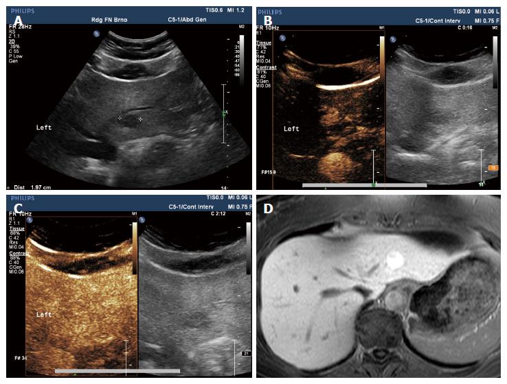
Figure 4 Focal nodular hyperplasia.
Female, 52 years of age, with anaemia. Native ultrasound (A) shows 19 mm hypoechoic lesion in segment S2 of the left liver lobe. On subsequent contrast-enhanced ultrasonography (CEUS) examination, there is increased centrifugal perfusion in arterial phase (B); In the late phase (C), the lesion blends in with the surrounding liver parenchyma and there is no sign of washout. The CEUS diagnosis was FNH. Magnetic resonance imaging (MRI); (D) performed for another reason after three months verifies the diagnosis - lesion with high signal on hepatobiliary phase of contrast-enhanced MRI.
- Citation: Smajerova M, Petrasova H, Little J, Ovesna P, Andrasina T, Valek V, Nemcova E, Miklosova B. Contrast-enhanced ultrasonography in the evaluation of incidental focal liver lesions: A cost-effectiveness analysis. World J Gastroenterol 2016; 22(38): 8605-8614
- URL: https://www.wjgnet.com/1007-9327/full/v22/i38/8605.htm
- DOI: https://dx.doi.org/10.3748/wjg.v22.i38.8605


