Copyright
©The Author(s) 2016.
World J Gastroenterol. Oct 14, 2016; 22(38): 8605-8614
Published online Oct 14, 2016. doi: 10.3748/wjg.v22.i38.8605
Published online Oct 14, 2016. doi: 10.3748/wjg.v22.i38.8605
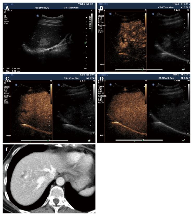
Figure 3 Haemangioma.
Female, 57 years of age. Ultrasound scan performed for dyspepsia, a hyperechoic lesion found in segment S8 of the right liver lobe (A); Peripheral nodular enhancement after application of the contrast agent (B, C); In the late phase, the lesion blends in with the surrounding liver parenchyma (D); An identical pattern of enhancement could be seen in CT performed later from a different indication (E). Both findings suggest haemangioma.
- Citation: Smajerova M, Petrasova H, Little J, Ovesna P, Andrasina T, Valek V, Nemcova E, Miklosova B. Contrast-enhanced ultrasonography in the evaluation of incidental focal liver lesions: A cost-effectiveness analysis. World J Gastroenterol 2016; 22(38): 8605-8614
- URL: https://www.wjgnet.com/1007-9327/full/v22/i38/8605.htm
- DOI: https://dx.doi.org/10.3748/wjg.v22.i38.8605


