Copyright
©The Author(s) 2016.
World J Gastroenterol. Oct 14, 2016; 22(38): 8459-8471
Published online Oct 14, 2016. doi: 10.3748/wjg.v22.i38.8459
Published online Oct 14, 2016. doi: 10.3748/wjg.v22.i38.8459
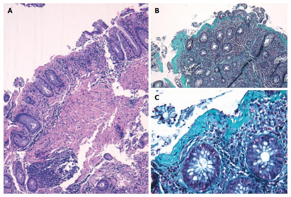
Figure 2 Photomicrographs of a colonic specimen from a collagenous colitis patient showing detachment of superficial epithelium.
Hematoxylin-eosin staining, magnification × 100 (A) and thick subepithelial collagen band, Gomori’s Trichrome staining (B and C, magnification × 100, × 400, respectively).
- Citation: Guagnozzi D, Landolfi S, Vicario M. Towards a new paradigm of microscopic colitis: Incomplete and variant forms. World J Gastroenterol 2016; 22(38): 8459-8471
- URL: https://www.wjgnet.com/1007-9327/full/v22/i38/8459.htm
- DOI: https://dx.doi.org/10.3748/wjg.v22.i38.8459


