Copyright
©The Author(s) 2016.
World J Gastroenterol. Oct 7, 2016; 22(37): 8439-8446
Published online Oct 7, 2016. doi: 10.3748/wjg.v22.i37.8439
Published online Oct 7, 2016. doi: 10.3748/wjg.v22.i37.8439
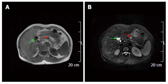
Figure 3 Magnetic resonance imaging findings.
A: The mass in the pancreatic body (red arrow) and gallbladder (green arrow) appeared hypointense on T1 weighted images; B: The mass in the pancreatic body (red arrow) appeared inhomogeneously hyperintense and the enlarged gallbladder (green arrow) appeared hyperintense on T2 weighted images.
- Citation: Xu SY, Sun K, Owusu-Ansah KG, Xie HY, Zhou L, Zheng SS, Wang WL. Central pancreatectomy for pancreatic schwannoma: A case report and literature review. World J Gastroenterol 2016; 22(37): 8439-8446
- URL: https://www.wjgnet.com/1007-9327/full/v22/i37/8439.htm
- DOI: https://dx.doi.org/10.3748/wjg.v22.i37.8439


