Copyright
©The Author(s) 2016.
World J Gastroenterol. Oct 7, 2016; 22(37): 8349-8360
Published online Oct 7, 2016. doi: 10.3748/wjg.v22.i37.8349
Published online Oct 7, 2016. doi: 10.3748/wjg.v22.i37.8349
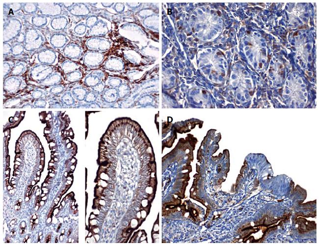
Figure 5 Gastric mucosa samples stained with CD10.
A: In patients with gastritis, diffuse CD10 positivity is shown in lymphocytes under the surface epithelium. Staining was not observed in the glandular epithelium (CD10, magnification × 200); B: CD10 positivity is shown in lymphocytes inside the epithelium of patients with gastritis. Staining was not observed in the glandular epithelium (CD10, magnification × 400); C: Intense positive staining is shown in the brush borders of the surface epithelium and supranuclear cytoplasm from healthy duodenal samples (CD10, magnification × 100 and 200); D: Loss of CD10 staining on the surface epithelium of duodenal samples from patients with duodenitis.
- Citation: Islek A, Yilmaz A, Elpek GO, Erin N. Childhood chronic gastritis and duodenitis: Role of altered sensory neuromediators. World J Gastroenterol 2016; 22(37): 8349-8360
- URL: https://www.wjgnet.com/1007-9327/full/v22/i37/8349.htm
- DOI: https://dx.doi.org/10.3748/wjg.v22.i37.8349


