Copyright
©The Author(s) 2016.
World J Gastroenterol. Oct 7, 2016; 22(37): 8334-8348
Published online Oct 7, 2016. doi: 10.3748/wjg.v22.i37.8334
Published online Oct 7, 2016. doi: 10.3748/wjg.v22.i37.8334
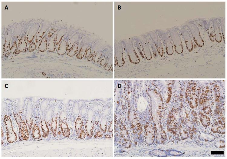
Figure 5 Immunohistochemical detection of Ki67 in Winnie distal colonic mucosa.
A: Immunostaining of Ki67 in the distal colon of untreated C57BL6 mice. Image representative of Ki67 localisation in the distal colon of the four C57BL6 mice examined; B: Distal colonic Ki67 localisation representative of six C57BL6 mice exposed to three cycles of 1% dextran sulphate sodium (DSS); C: Distal colon of Winnie mouse without exposure to three cycles of DSS. Ki67-labelling in the epithelium is visible apically approximately half the crypt length; D: Ki67 immunolabelling of the Winnie distal colon exposed to three cycles of DSS. Crypt base proliferative zone extends approximately two-thirds of the crypt length. Submucosal gland (arrowhead) displays few positive nuclei. Scale bar represents a distance of 50 μm.
- Citation: Randall-Demllo S, Fernando R, Brain T, Sohal SS, Cook AL, Guven N, Kunde D, Spring K, Eri R. Characterisation of colonic dysplasia-like epithelial atypia in murine colitis. World J Gastroenterol 2016; 22(37): 8334-8348
- URL: https://www.wjgnet.com/1007-9327/full/v22/i37/8334.htm
- DOI: https://dx.doi.org/10.3748/wjg.v22.i37.8334


