Copyright
©The Author(s) 2016.
World J Gastroenterol. Oct 7, 2016; 22(37): 8334-8348
Published online Oct 7, 2016. doi: 10.3748/wjg.v22.i37.8334
Published online Oct 7, 2016. doi: 10.3748/wjg.v22.i37.8334
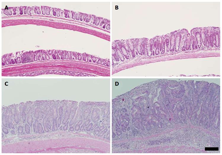
Figure 3 Distal colonic mucosal alterations induced by three cycles of dextran sulphate sodium.
A: Representative image of the distal colon obtained from one of four untreated C57BL/6 mouse; B: C57BL/6 distal colon exposed to dextran sulphate sodium (DSS). Image representative of six eighteen week-old C57BL/6 mice exposed to three cycles of 1% DSS; C: Distal colon of Winnie mouse without exposure to DSS. Crypts are hyperplastic and the mucosa displays a marked leukocytic infiltration. Image representative of six untreated Winnie mice examined; D: Distal colon of Winnie mouse displaying colonic hyperplasia and mild focal dysplasia following three cycles of 1% DSS. Mucosa displays features of an active chronic inflammation, with prominent submucosal leukocytic infiltrate. Extensive crypt hyperplasia is visible with a focus of atypical glandular architecture (asterisk). Note the loss of surface epithelium. Numerous mitotic figures are evident. Image representative of the hyperplasia and dysplastic foci total of eleven Winnie mice exposed to three cycles of 1% DSS. All sections stained with HE, scale bar is equivalent to 100 μm.
- Citation: Randall-Demllo S, Fernando R, Brain T, Sohal SS, Cook AL, Guven N, Kunde D, Spring K, Eri R. Characterisation of colonic dysplasia-like epithelial atypia in murine colitis. World J Gastroenterol 2016; 22(37): 8334-8348
- URL: https://www.wjgnet.com/1007-9327/full/v22/i37/8334.htm
- DOI: https://dx.doi.org/10.3748/wjg.v22.i37.8334


