Copyright
©The Author(s) 2016.
World J Gastroenterol. Sep 28, 2016; 22(36): 8242-8246
Published online Sep 28, 2016. doi: 10.3748/wjg.v22.i36.8242
Published online Sep 28, 2016. doi: 10.3748/wjg.v22.i36.8242
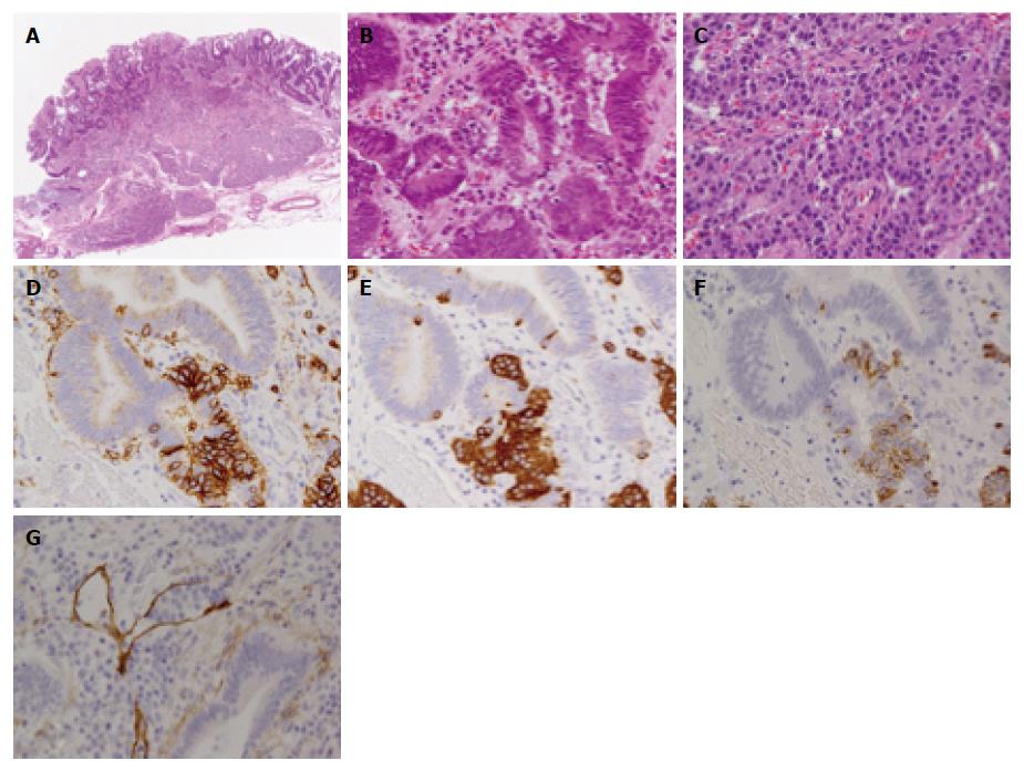
Figure 3 Histopathological results of the resected specimen.
A: Low power view of a histological section demonstrates that the neuroendocrine tumor (NET) in the submucosa was covered with adenocarcinoma (HE stain × 40); B: Both component cells had mixed in some parts and formed a tumor alveolar (H&E stain × 400); C: High power view of NET in submucosa (HE stain × 400); D: CD56; E: syNaptophisin; F: Chromogranin A staining were positive in the NET cells; G: Lymphatic invasion of the NET component seen with D2-40 staining (× 400).
- Citation: Kaneko H, Miyake A, Ishii Y, Sue S, Miwa H, Sasaki T, Tamura T, Kondo M, Maeda S. Case of a tumor comprising gastric cancer and duodenal neuroendocrine tumor. World J Gastroenterol 2016; 22(36): 8242-8246
- URL: https://www.wjgnet.com/1007-9327/full/v22/i36/8242.htm
- DOI: https://dx.doi.org/10.3748/wjg.v22.i36.8242


