Copyright
©The Author(s) 2016.
World J Gastroenterol. Sep 28, 2016; 22(36): 8234-8241
Published online Sep 28, 2016. doi: 10.3748/wjg.v22.i36.8234
Published online Sep 28, 2016. doi: 10.3748/wjg.v22.i36.8234
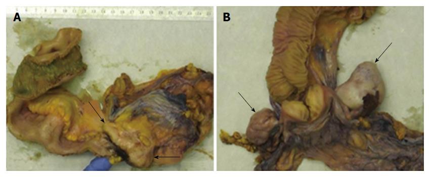
Figure 5 Gross pathology of the surgical specimens.
A: Gross image showing a tumor mass (arrows) that involves the appendix, the ileocecal valve and the cecum. Surgical resection was performed after eradication of amebiasis and chemotherapy. B: Gross image of the Douglas-resection showing bilateral ovary metastases (arrows).
- Citation: Grosse A. Diagnosis of colonic amebiasis and coexisting signet-ring cell carcinoma in intestinal biopsy. World J Gastroenterol 2016; 22(36): 8234-8241
- URL: https://www.wjgnet.com/1007-9327/full/v22/i36/8234.htm
- DOI: https://dx.doi.org/10.3748/wjg.v22.i36.8234


