Copyright
©The Author(s) 2016.
World J Gastroenterol. Sep 28, 2016; 22(36): 8234-8241
Published online Sep 28, 2016. doi: 10.3748/wjg.v22.i36.8234
Published online Sep 28, 2016. doi: 10.3748/wjg.v22.i36.8234
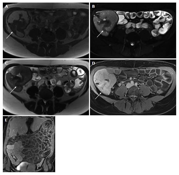
Figure 1 Magnetic resonance imaging showed mural thickening of the ileocecum and the appendix, which was interpreted as being inflammatory in nature.
A-E: Magnetic resonance images (A: T2-weighted magnetization transfer contrast transversal; B: T2-weighted fat saturated transversal; C: T2-weighted half-Fourier acquired single-shot turbo spin-echo transversal; D: T1-weighted VIBE-Dixon postcontrast transversal, E: T1-weighted VIBE-Dixon postcontrast coronal) showing mural thickening (arrows in A-C) of the ileocecum and the appendix with contrast enhancement (arrow in D, asterix in E).
- Citation: Grosse A. Diagnosis of colonic amebiasis and coexisting signet-ring cell carcinoma in intestinal biopsy. World J Gastroenterol 2016; 22(36): 8234-8241
- URL: https://www.wjgnet.com/1007-9327/full/v22/i36/8234.htm
- DOI: https://dx.doi.org/10.3748/wjg.v22.i36.8234


