Copyright
©The Author(s) 2016.
World J Gastroenterol. Sep 28, 2016; 22(36): 8203-8210
Published online Sep 28, 2016. doi: 10.3748/wjg.v22.i36.8203
Published online Sep 28, 2016. doi: 10.3748/wjg.v22.i36.8203
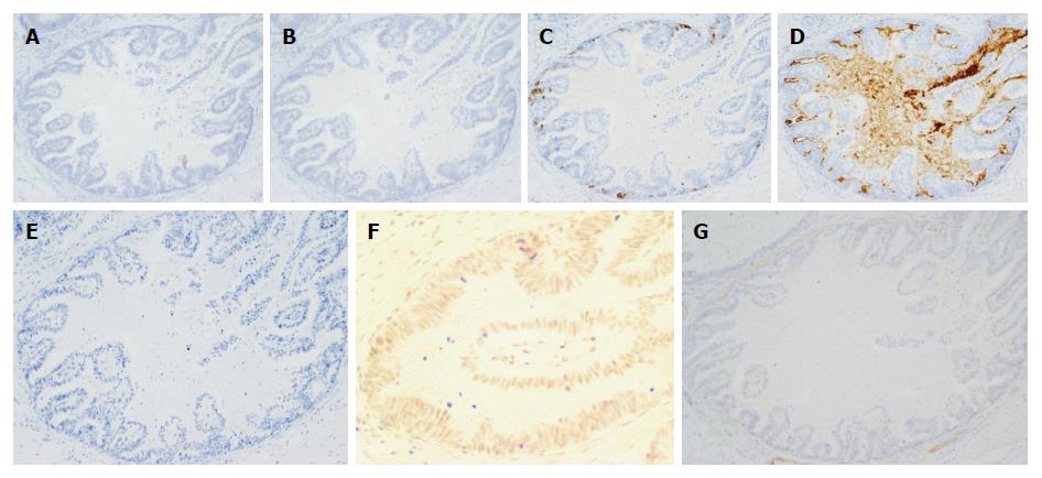
Figure 3 Immunohistochemical examination of resected specimens (case 1).
A: MUC2; B: MUC5AC; C: MUC6; D: CD10; E: AFP; F: Glypican 3; G: SALL 4. The lesion had focal positivity for MUC6, diffuse positivity for CD10, weak positivity for Glypican 3 and negative staining for MUC2, MUC5AC, AFP and SALL4.
- Citation: Matsumoto K, Ueyama H, Matsumoto K, Akazawa Y, Komori H, Takeda T, Murakami T, Asaoka D, Hojo M, Tomita N, Nagahara A, Kajiyama Y, Yao T, Watanabe S. Clinicopathological features of alpha-fetoprotein producing early gastric cancer with enteroblastic differentiation. World J Gastroenterol 2016; 22(36): 8203-8210
- URL: https://www.wjgnet.com/1007-9327/full/v22/i36/8203.htm
- DOI: https://dx.doi.org/10.3748/wjg.v22.i36.8203


