Copyright
©The Author(s) 2016.
World J Gastroenterol. Sep 28, 2016; 22(36): 8203-8210
Published online Sep 28, 2016. doi: 10.3748/wjg.v22.i36.8203
Published online Sep 28, 2016. doi: 10.3748/wjg.v22.i36.8203
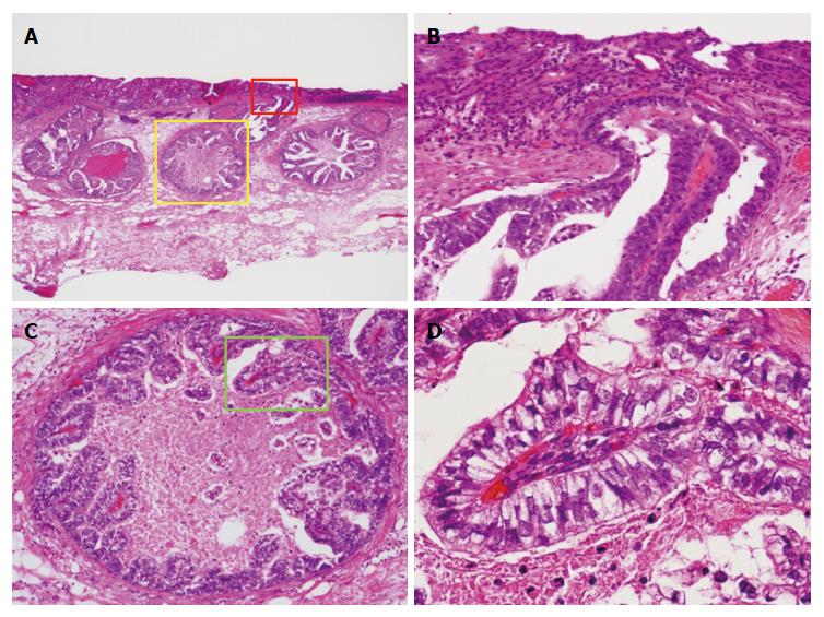
Figure 2 Histological examination of the resected specimens with hematoxylin and eosin stain (case 1).
A and B: The superficial mucosal layer was covered with a well differentiated adenocarcinoma; B-D: Carcinoma cells with clear cytoplasm had tubulopapillary growth, resembling the primitive gut. These carcinoma cells arose from the deeper part of the mucosal layer and invaded into the submucosal layer by venous invasion. There was no severe stromal reaction at the submucosal layer. Pathological diagnosis was as follows: stomach (ESD): adenocarcinoma with enteroblastic differentiation, 0-IIc, 11 mm × 9 mm, well-differentiated carcinoma > moderately-differentiated carcinoma, papillary adenocarcinoma, SM (1500 μm), ly (-), v (+), HM0, VM0. SM: Submucosa; ESD: Endoscopic submucosal dissection.
- Citation: Matsumoto K, Ueyama H, Matsumoto K, Akazawa Y, Komori H, Takeda T, Murakami T, Asaoka D, Hojo M, Tomita N, Nagahara A, Kajiyama Y, Yao T, Watanabe S. Clinicopathological features of alpha-fetoprotein producing early gastric cancer with enteroblastic differentiation. World J Gastroenterol 2016; 22(36): 8203-8210
- URL: https://www.wjgnet.com/1007-9327/full/v22/i36/8203.htm
- DOI: https://dx.doi.org/10.3748/wjg.v22.i36.8203


