Copyright
©The Author(s) 2016.
World J Gastroenterol. Sep 21, 2016; 22(35): 7973-7982
Published online Sep 21, 2016. doi: 10.3748/wjg.v22.i35.7973
Published online Sep 21, 2016. doi: 10.3748/wjg.v22.i35.7973
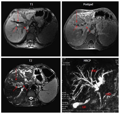
Figure 4 Magnetic resonance imaging with Magnetic retrograde cholangiogram in a patient with portal biliopathy.
T1 weighted, T2 weighted and post-gadolinium (postgad) images. Mass lesion (short arrow) is seen at the level of porta hepatis with dilated bile ducts (long arrow) and signal void (curved arrow), depicting collateral channels compressing bile ducts, MRCP. Common bile duct (CBD) shows luminal irregularity. There is a localized irregular stricture in common hepatic duct with upstream gross dilatation of intrahepatic ducts (IHD).
- Citation: Khuroo MS, Rather AA, Khuroo NS, Khuroo MS. Portal biliopathy. World J Gastroenterol 2016; 22(35): 7973-7982
- URL: https://www.wjgnet.com/1007-9327/full/v22/i35/7973.htm
- DOI: https://dx.doi.org/10.3748/wjg.v22.i35.7973


