Copyright
©The Author(s) 2016.
World J Gastroenterol. Sep 21, 2016; 22(35): 7973-7982
Published online Sep 21, 2016. doi: 10.3748/wjg.v22.i35.7973
Published online Sep 21, 2016. doi: 10.3748/wjg.v22.i35.7973
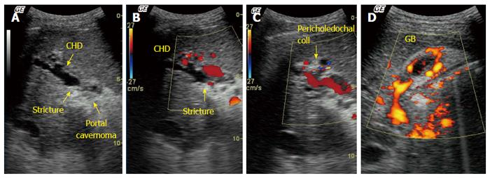
Figure 3 Ultrasound with Doppler in a patient with extrahepatic portal venous obstruction and portal biliopathy.
A: Gray scale image shows an echogenic mass in hilum (portal cavernoma). Common hepatic duct (CHD) shows dilatation and luminal irregularity with a stricture at the level of the cavernoma; B: Color Doppler image showed multiple pericholedochal collaterals around the strictured bile duct (arrow); C: Color Doppler image shows pericholedochal collaterals and recanalized irregular portal vein; D: Power Doppler images of gall bladder. Gall bladder wall is thickened with multiple collaterals within the gall bladder wall.
- Citation: Khuroo MS, Rather AA, Khuroo NS, Khuroo MS. Portal biliopathy. World J Gastroenterol 2016; 22(35): 7973-7982
- URL: https://www.wjgnet.com/1007-9327/full/v22/i35/7973.htm
- DOI: https://dx.doi.org/10.3748/wjg.v22.i35.7973


