Copyright
©The Author(s) 2016.
World J Gastroenterol. Sep 21, 2016; 22(35): 7973-7982
Published online Sep 21, 2016. doi: 10.3748/wjg.v22.i35.7973
Published online Sep 21, 2016. doi: 10.3748/wjg.v22.i35.7973
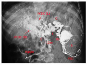
Figure 1 Splenoportovenography in a patient with extrahepatic portal venous obstruction and portal cavernoma.
Contrast within splenic pulp (S) is drained by multiple splenic vein channels (SV) into large peripancreatic collaterals (C). There are two broad parallel conglomerate of veins in the porta hepatis (arrows) formed by right and left paracholedochal collaterals [PCDC (R) and PCDC (L) respectively], forming the portal cavernoma. Main portal vein is not seen (thrombosed). Retrograde filling of perisplenic collaterals (PSC), gastroduodenal vein (GDV) and posterior-superior pancreaticoduodenal vein (PSPDV) is seen.
- Citation: Khuroo MS, Rather AA, Khuroo NS, Khuroo MS. Portal biliopathy. World J Gastroenterol 2016; 22(35): 7973-7982
- URL: https://www.wjgnet.com/1007-9327/full/v22/i35/7973.htm
- DOI: https://dx.doi.org/10.3748/wjg.v22.i35.7973


