Copyright
©The Author(s) 2016.
World J Gastroenterol. Sep 14, 2016; 22(34): 7797-7805
Published online Sep 14, 2016. doi: 10.3748/wjg.v22.i34.7797
Published online Sep 14, 2016. doi: 10.3748/wjg.v22.i34.7797
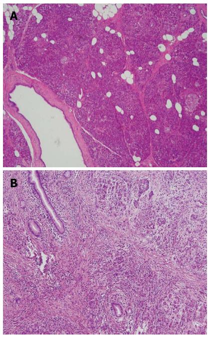
Figure 1 Photomicrographs of the pathological examination for pancreatic fibrosis.
A: No significant fibrosis; B: Severe fibrosis (hematoxylin-eosin staining; original magnification, × 100).
- Citation: Hu BY, Wan T, Zhang WZ, Dong JH. Risk factors for postoperative pancreatic fistula: Analysis of 539 successive cases of pancreaticoduodenectomy. World J Gastroenterol 2016; 22(34): 7797-7805
- URL: https://www.wjgnet.com/1007-9327/full/v22/i34/7797.htm
- DOI: https://dx.doi.org/10.3748/wjg.v22.i34.7797


