Copyright
©The Author(s) 2016.
World J Gastroenterol. Sep 7, 2016; 22(33): 7536-7558
Published online Sep 7, 2016. doi: 10.3748/wjg.v22.i33.7536
Published online Sep 7, 2016. doi: 10.3748/wjg.v22.i33.7536
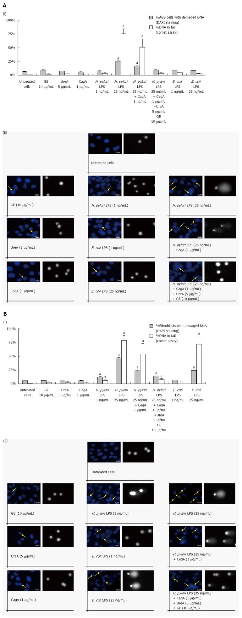
Figure 6 Genotoxic properties of Helicobacter pylori antigens assessed by 4’,6-diamidino-2-phenylindole staining and a comet assay.
The influence of Helicobacter pylori or E. coli antigens on DNA stability was estimated in AGS cells (A) and fibroblasts (B) 24 h after the challenge. A 4’,6-diamidino-2-phenylindole (DAPI) staining assay was used to visualize DNA changes in cell nuclei and a comet assay was applied to confirm DNA damage by the measurement of the percentage of DNA in the comet tail. Mean values were replicated of 50 comets each. The values are the means ± SD. aP < 0.05 vs untreated cells. (i) the graphs indicate the percentage of cells with DAPI stained nuclei (blue bars) and the percentage of nuclei with DNA in the comet tail (grey bars); and (ii) visualization of morphological changes in the cell nuclei after the treatment with bacterial antigens followed by DAPI staining and a comet assay. Imaging was performed using a fluorescent microscope (Axio Scope A1, Zeiss, Germany). The arrows indicate the damaged cell nuclei (magnification, × 1000). DNA tails were measured using the CASP software (latest beta version 1.2.3.beta2). Representative results of the comet assay were selected (magnification, × 400).
- Citation: Mnich E, Kowalewicz-Kulbat M, Sicińska P, Hinc K, Obuchowski M, Gajewski A, Moran AP, Chmiela M. Impact of Helicobacter pylori on the healing process of the gastric barrier. World J Gastroenterol 2016; 22(33): 7536-7558
- URL: https://www.wjgnet.com/1007-9327/full/v22/i33/7536.htm
- DOI: https://dx.doi.org/10.3748/wjg.v22.i33.7536


