Copyright
©The Author(s) 2016.
World J Gastroenterol. Aug 21, 2016; 22(31): 7157-7165
Published online Aug 21, 2016. doi: 10.3748/wjg.v22.i31.7157
Published online Aug 21, 2016. doi: 10.3748/wjg.v22.i31.7157
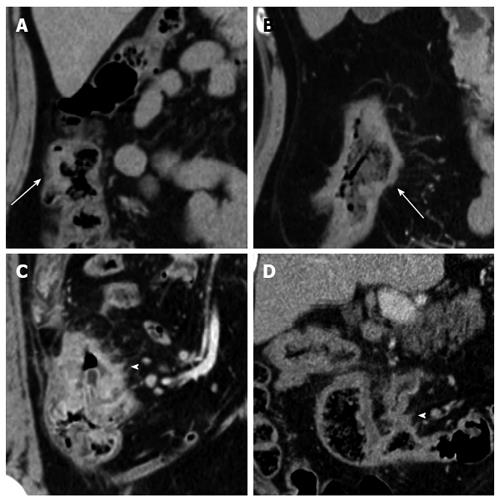
Figure 1 Extramural vascular invasion status as detected on multiple-row detector computed tomography in colon cancer.
A: A negative EMVI score of 0 on coronal MDCT. The tumor was located within the surface of the colon facing the retroperitoneum; there was no adjacent vessel (arrow). There was no indication of EMVI; B: A negative EMVI score of 1 on a coronal MDCT. The tumor was located within the mesenteric side of the colon, but no vascular invasion was seen (arrow). The image was suspicious for EMVI with a confidence level < 50%; C: A positive EMVI score of 2 on a coronal MDCT. The involved vein was small and slightly dilated (arrow head). The image was suspicious for EMVI with a confidence level > 50%; D: A positive EMVI score of 3 on an axial MDCT. The involved vein was grossly dilated (arrow head). There was a definite indication of EMVI. EMVI: Extramural vascular invasion; MDCT: Multiple-row detector computed tomography.
- Citation: Yao X, Yang SX, Song XH, Cui YC, Ye YJ, Wang Y. Prognostic significance of computed tomography-detected extramural vascular invasion in colon cancer. World J Gastroenterol 2016; 22(31): 7157-7165
- URL: https://www.wjgnet.com/1007-9327/full/v22/i31/7157.htm
- DOI: https://dx.doi.org/10.3748/wjg.v22.i31.7157


