Copyright
©The Author(s) 2016.
World J Gastroenterol. Aug 14, 2016; 22(30): 6960-6964
Published online Aug 14, 2016. doi: 10.3748/wjg.v22.i30.6960
Published online Aug 14, 2016. doi: 10.3748/wjg.v22.i30.6960
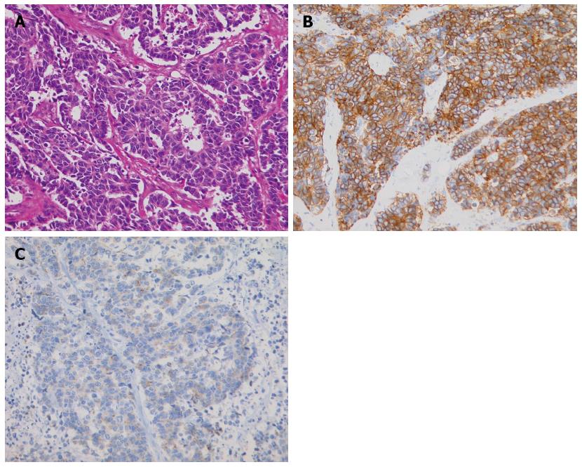
Figure 4 Histopathologic appearance of the neuroendocrine carcinoma.
A: The cells were round or oval, hyperchromatic, and had an increased nucleus-to-cytoplasm ratio (hematoxylin-eosin staining, magnification × 200); B: Immunohistochemically, tumor cells were diffusely positive for CD56, a membrane protein usually present in neuroendocrine cells; C: Immunohistochemically, tumor cells were positive for synaptophysin, which is typically expressed on the surface of neurons or endothelial cells.
- Citation: Oshiro Y, Gen R, Hashimoto S, Oda T, Sato T, Ohkohchi N. Neuroendocrine carcinoma of the extrahepatic bile duct: A case report. World J Gastroenterol 2016; 22(30): 6960-6964
- URL: https://www.wjgnet.com/1007-9327/full/v22/i30/6960.htm
- DOI: https://dx.doi.org/10.3748/wjg.v22.i30.6960


