Copyright
©The Author(s) 2016.
World J Gastroenterol. Aug 14, 2016; 22(30): 6955-6959
Published online Aug 14, 2016. doi: 10.3748/wjg.v22.i30.6955
Published online Aug 14, 2016. doi: 10.3748/wjg.v22.i30.6955
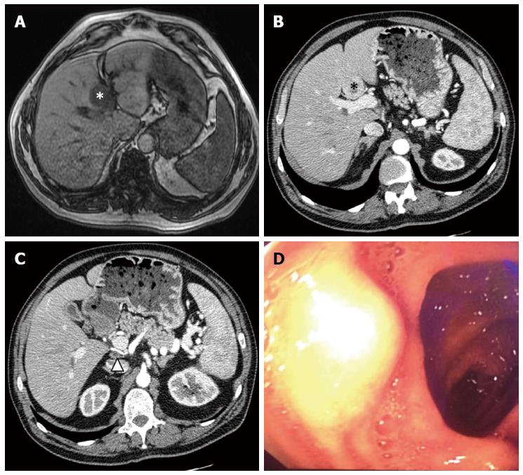
Figure 1 Pre-operative imaging.
A: Pre-operative MRI of the liver, showing liver metastases of gastrinoma (white star); B: Pre-operative CT scan of the abdomen, showing liver metastases of gastrinoma (black star); C: Pre-operative CT scan of the abdomen, showing the retroportal lymph node (black triangle); D: Upper endoscopy, showing the tumor on the superior duodenal flexure. MRI: Magnetic resonance imaging; CT: Computed tomography.
- Citation: Gracient A, Rebibo L, Delcenserie R, Yzet T, Regimbeau JM. Combined radiologic and endoscopic treatment (using the “rendezvous technique”) of a biliary fistula following left hepatectomy. World J Gastroenterol 2016; 22(30): 6955-6959
- URL: https://www.wjgnet.com/1007-9327/full/v22/i30/6955.htm
- DOI: https://dx.doi.org/10.3748/wjg.v22.i30.6955


