Copyright
©The Author(s) 2016.
World J Gastroenterol. Aug 14, 2016; 22(30): 6817-6828
Published online Aug 14, 2016. doi: 10.3748/wjg.v22.i30.6817
Published online Aug 14, 2016. doi: 10.3748/wjg.v22.i30.6817
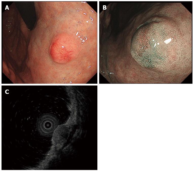
Figure 1 Gastric neuroendocrine tumor.
A: Conventional endoscopic image with white light demonstrates a hemispherical reddish submucosal tumor; B: Magnifying endoscopic image with narrow band imaging demonstrates gastric pit structures present on the surface of the tumor; C: Endoscopic ultrasound (EUS, 20 MHz) demonstrates a hypoechoic intramural structure in the second layer, which corresponds to the submucosal layer of the gastric wall. EUS: Endoscopic ultrasonography.
- Citation: Sato Y, Hashimoto S, Mizuno KI, Takeuchi M, Terai S. Management of gastric and duodenal neuroendocrine tumors. World J Gastroenterol 2016; 22(30): 6817-6828
- URL: https://www.wjgnet.com/1007-9327/full/v22/i30/6817.htm
- DOI: https://dx.doi.org/10.3748/wjg.v22.i30.6817


