Copyright
©The Author(s) 2016.
World J Gastroenterol. Jan 21, 2016; 22(3): 917-932
Published online Jan 21, 2016. doi: 10.3748/wjg.v22.i3.917
Published online Jan 21, 2016. doi: 10.3748/wjg.v22.i3.917
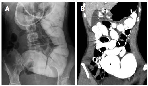
Figure 2 Computed tomography enteroclysis depiction of small bowel stricture.
Fluoroscopic (A) and computed tomography (B) images from an enteroclysis demonstrating a 9 cm stricture in the mid small bowel (arrows) with proximal small bowel dilation (asterisks), which was confirmed during subsequent surgical resection.
- Citation: Kilcoyne A, Kaplan JL, Gee MS. Inflammatory bowel disease imaging: Current practice and future directions. World J Gastroenterol 2016; 22(3): 917-932
- URL: https://www.wjgnet.com/1007-9327/full/v22/i3/917.htm
- DOI: https://dx.doi.org/10.3748/wjg.v22.i3.917


