Copyright
©The Author(s) 2016.
World J Gastroenterol. Jul 14, 2016; 22(26): 6036-6048
Published online Jul 14, 2016. doi: 10.3748/wjg.v22.i26.6036
Published online Jul 14, 2016. doi: 10.3748/wjg.v22.i26.6036
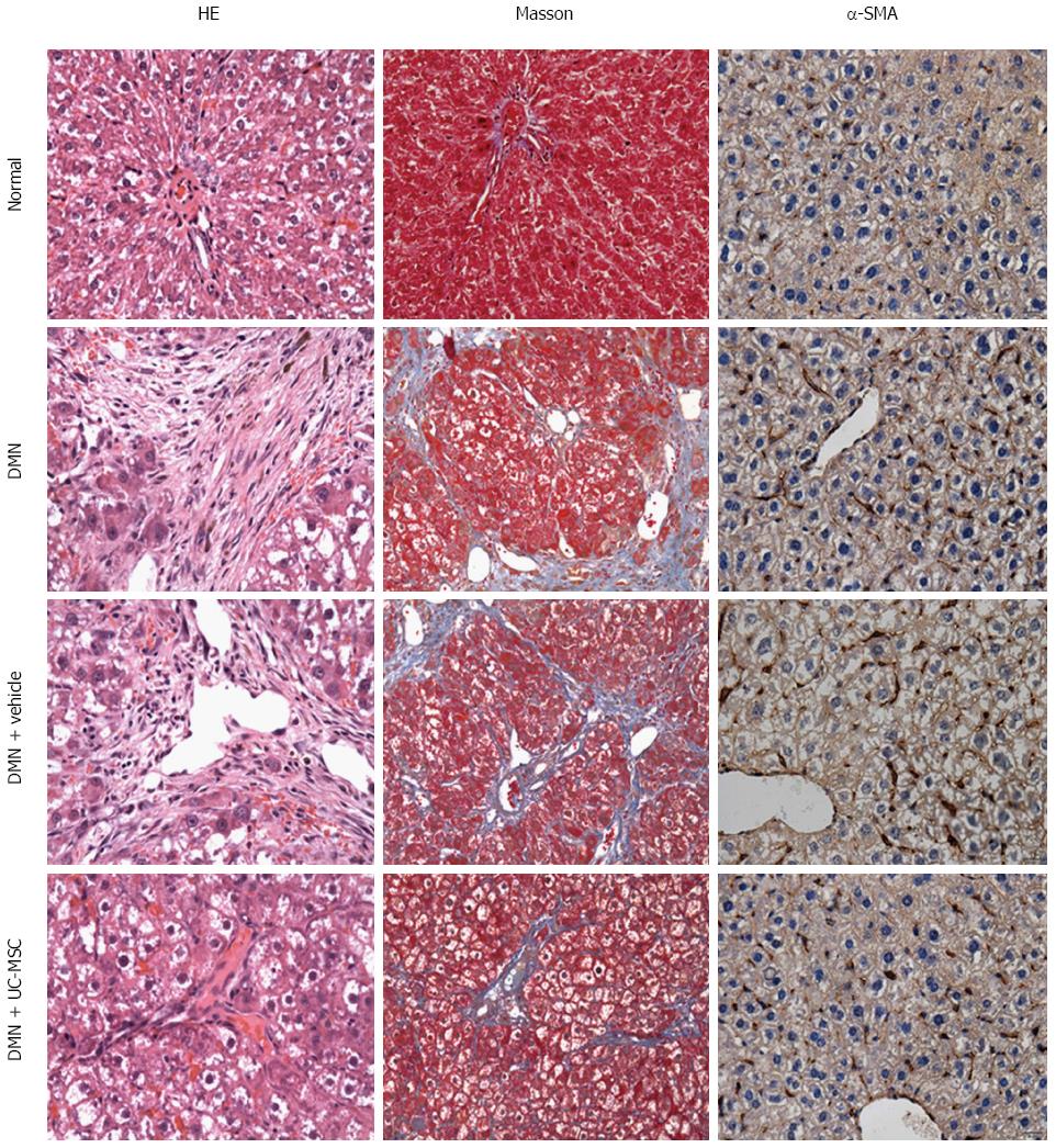
Figure 2 Umbilical cord-derived mesenchymal stem cells transplantation alleviates liver fibrosis.
Hematoxylin and eosin (HE) staining of liver sections showing massive vacuolar degeneration of hepatocytes and intense neutrophil infiltration in DMN-treated rats and control rats (first column, HE staining). Masson’s trichrome staining showed periportal fibrosis with fibrous septa extending into adjacent portal tracts and the terminal hepatic venule in DMN-treated rats and DMN + Vehicle animals (second column, Masson staining), suggesting the successful establishment of the animal model of hepatic fibrosis. Liver fibrosis and inflammation were significantly decreased by transfusion with UC-MSCs in UC-MSC-treated rats compared with the DMN + Vehicle group, which was characterized by short septa extending into lobules or portal-portal septa (first column, HE staining, and second column, Masson staining). Alpha-SMA is a marker of activated hepatic stellate cells, which plays key roles in the pathogenesis of liver fibrosis. Increased expression of α-SMA was observed in the model and DMN + Vehicle groups, and was decreased by UC-MSCs transfusion (third column, α-SMA immunohistochemical staining).
- Citation: Chai NL, Zhang XB, Chen SW, Fan KX, Linghu EQ. Umbilical cord-derived mesenchymal stem cells alleviate liver fibrosis in rats. World J Gastroenterol 2016; 22(26): 6036-6048
- URL: https://www.wjgnet.com/1007-9327/full/v22/i26/6036.htm
- DOI: https://dx.doi.org/10.3748/wjg.v22.i26.6036


