Copyright
©The Author(s) 2016.
World J Gastroenterol. Jun 28, 2016; 22(24): 5598-5608
Published online Jun 28, 2016. doi: 10.3748/wjg.v22.i24.5598
Published online Jun 28, 2016. doi: 10.3748/wjg.v22.i24.5598
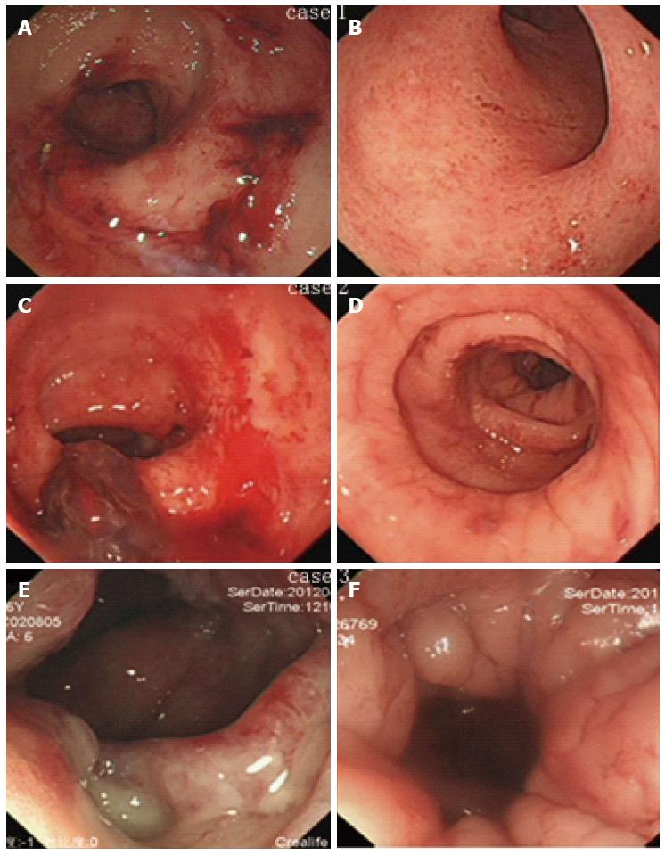
Figure 3 Classical endoscopic images before colostomy and at stoma reversal.
Severe active bleeding (A and C), or confluent telangiectasia, edema and ulcer (E) were observed in cases 1-3 before colostomy, while these lesions was greatly improved at stoma reversal (B, D, F).
- Citation: Yuan ZX, Ma TH, Wang HM, Zhong QH, Yu XH, Qin QY, Wang JP, Wang L. Colostomy is a simple and effective procedure for severe chronic radiation proctitis. World J Gastroenterol 2016; 22(24): 5598-5608
- URL: https://www.wjgnet.com/1007-9327/full/v22/i24/5598.htm
- DOI: https://dx.doi.org/10.3748/wjg.v22.i24.5598


