Copyright
©The Author(s) 2016.
World J Gastroenterol. Jun 28, 2016; 22(24): 5479-5494
Published online Jun 28, 2016. doi: 10.3748/wjg.v22.i24.5479
Published online Jun 28, 2016. doi: 10.3748/wjg.v22.i24.5479
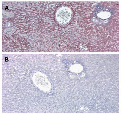
Figure 7 Immune labeling of CTT liver by anti fibulin-5 monoclonal antibody (A) and corresponding negative control (B).
A: A central vein is seen in the upper center and a portal triad to the right. The dark red staining denotes the distribution of the antibody which is particularly intense surround the central vein. This is a low power magnification; B: In the negative control no staining is evident.
- Citation: Tobi M, Thomas P, Ezekwudo D. Avoiding hepatic metastasis naturally: Lessons from the cotton top tamarin (Saguinus oedipus). World J Gastroenterol 2016; 22(24): 5479-5494
- URL: https://www.wjgnet.com/1007-9327/full/v22/i24/5479.htm
- DOI: https://dx.doi.org/10.3748/wjg.v22.i24.5479


